HEMOGLOBIN SYNTHESIS, FUNCTION, CATABOLISM AND EXCREATION
We know well that the main function of the red blood cell is to carry oxygen to the tissues and return carbon dioxide to the lungs. This process of gaseous exchange is achieved by the specialized protein in the red blood cell, hemoglobin. Each red blood cell then contains approximately 640 million hemoglobin molecules.

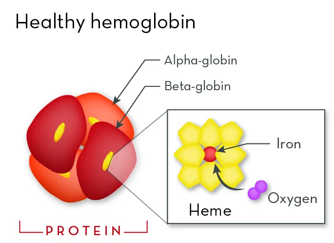
Hemoglobin is an oxygen-binding protein that is mainly found in erythrocytes that aids in transporting oxygen from the lungs to the tissues. The hemoglobins have a molecular weight of about 68,000 and compromises almost one third of the weight of a red blood cell. Each hemoglobin molecule is a tetramer made of four polypeptide globin chains. A heme moiety made up of an organic protoporphyrin ring and a central iron ion in the ferrous state(Fe2+) is found in each globin subunit. Each heme moiety contains an iron molecule that is able to bind and unbind oxygen, enabling the organism to transport oxygen. The most common type of hemoglobin in the adult is HbA, which comprises two alpha-globin and two beta-globin subunits. Different globin genes encode each type of globin subunit.
The two main components for haemoglobin synthesis are globin production and heme synthesis. The synthesis of haem and globin occurs separately. Haem is synthesized largely in the mitochondria, while globin is synthesized in the polyribosomes. Although the synthesis of these two components occur separately within developing red cell precursors, the rates of their synthesis are carefully coordinated for optimal efficiency of the haemoglobin assembly.
Red Blood
Cells; of Haemoglobin, its Creation & Degradation
“Every few seconds, someone,
somewhere, needs blood”
– By Vanessa Manogaren

“A healthy outside starts from the
inside.” –
Robert Urich.
Indeed, how wise is the above adage of
old. A healthy person derives from a
healthy body, primarily the cells and more significantly the red blood cells.
For the body, blood is a necessary element that houses the body’s most vital
blood cells. It has a somewhat sticky texture and is literally thicker than
water. This writing aims to emphasize
the pivot role red blood cells have in maintaining a safe and healthy blood.
Red blood cells, also known as erythrocytes, are the blood's cellular
building blocks and the oxygen-carrying units that give blood its distinctive
colour. A mature human red blood cell has a dumbbell-like form in profile, is
tiny, spherical, and biconcave. As it passes through incredibly tiny blood
veins, the flexible cell takes on a bell form. Do you know that there are
about 640 million haemoglobin molecules in a single red blood cell? A
conventional FBC blood laboratory test
can determine the blood's haemoglobin
count. Males have a healthy count of 13-18g/Dl, whereas a healthy female has a
level of 12-15g/Dl. Hemoglobin, a protein that reversibly binds and moves
carbon dioxide and oxygen, is present in the cytoplasm of red blood cells.
Hemoglobin is a tetramer that consists of four protein subunits called globin
chains. Haem binds to the iron molecule in globin to transport
oxygen. Each haemoglobin molecule can carry up to four molecules of oxygen or
carbon dioxide since the iron serves the primary role in binding gases.

In the bone
marrow, the red cell develops in stages. From a hemocytoblast, a multipotent
cell in the mesenchyme, it becomes an erythroblast (normoblast). During the
course of two to five days of development, the erythroblast progressively
occupies with haemoglobin and loses its nucleus and mitochondria, the particles
in the cytoplasm that provide the cell's source of energy. Colony
Forming Unit – Erythroid
(CFU-E), an erythroid stem cell, is
produced during the early stages of hematopoiesis The process of
erythropoiesis, which is primarily fueled by the hormone erythropoietin,
officially starts at this point. For example, when the body tissues lack
oxygen, they stimulate the kidney to release the hormone erythropoietin, which
in turn increases the production of red blood cells in the bone marrow, and
thus increases oxygen delivery to the tissues. CFU-E cells are located in
erythroid islands in the bone marrow, where they multiply and develop into
mature erythrocytes.
Proerythroblasts, erythroblasts, reticulocytes, and
erythrocytes are among the cell generations developed during the
differentiation process. The cell, which eventually develops into a completely
formed red cell, is known as a reticulocyte in a late stage. Each subsequent
cell population is histologically closer to erythrocytes. Eight enzymes work
together to synthesize heme in humans. The red blood cell travels through the
body in a convoluted manner, passing through the heart twice as it transforms
from a deoxygenated blood cell to an oxygenated blood cell. After being
synthesized, the red blood cell begins its capillary journey to the heart.
Right now, the blood cell is oxygen-depleted. Now that it has reached the
heart's vena cava, the deoxygenated red blood cell is forced into the right
atrium. The blood cell is subsequently forced past the tricuspid valve and into
the right ventricle when the right atrium closes. The red blood cell is then
pushed through the semi-lunar by the contraction of the right ventricle. The
red blood cell gets to the lungs via the pulmonary artery after exiting the
heart. The deoxygenated red blood cell becomes an oxygenated blood cell when it
takes up oxygen there. The pulmonary vein leads the blood cell into the left
atrium as it travels back to the heart. Red blood cells carry oxygenated blood
around the body as they pass through the aorta and enter the kidneys, trunk,
and other lower limbs. Before they pass away, they normally live for 120 days.
And that's how the whole mechanism works! Despite the fact that it seems to
take a while, depending on the person's heart rate, the overall process only
takes a minute
or less.

Haemoglobin: a question of salvaging, metabolism, and redistribution across cell membranes

Red blood cells have a finite life
span, of approximately 120 days. Erythrocytes' cell membrane deteriorates with
time while they are in circulation. Macrophages phagocytose an aged or
unviable erythrocyte when they recognize its morphological blueprint. The
spleen is the main site of eryptosis, which is the removal of erythrocytes. A
physiological number of RBCs are ensured in a healthy body by the balance
between eryptosis and erythropoiesis.
The
death of the red blood cell after 120 days marks the beginning of haemoglobin
catabolism or breakdown. As you may know, the two most crucial parts of haemoglobin
within an erythrocyte are globin chains and iron-containing heme groups. These
elements are separated after being phagocytosed by macrophages. In parallel
with the iron being drawn from heme, the polypeptide globin chains are broken
down into amino acids, a protein building block that is then used again for
protein synthesis. Heme is broken down into biliverdin which is converted into
bilirubin, a
yellow, insoluble in water, and
extremely toxic end product. while iron is then transferred back to the liver
and stored there as ferritin by transferrin, a protein mediator. Transferrin
plays the role of a mediator akin to trafficking iron across the cell
membrane. Hence, the iron molecule is later transported to the bone marrow to
be employed in new cycles of erythropoiesis. Bilirubin is eliminated through
urine as urobilin and faeces as stercobilin after going through additional
changes in the liver and intestines.
For
a more comprehensible illustration, watch the video that is featured below.
In essence, the synthesis and catabolism of each
haemoglobin in the blood is a complex and extremely detailed-oriented process.
I would like to drive home the message that blood formation is an essential and
much needed aspect of the human body, as such, we should take every effort to
preserve this priceless entity and to champion it for safe blood donation, as
the demand for blood is critical and especially needed in surgeries.
Give
those who need it this indispensable gift of life!

Video: The Breakdown of Haemoglobin Made Simple
Adapted from, MedsXclusive Learning https://www.youtube.com/watch?v=pd-BTcu00nE
The Purpose
The need for safe blood is vast,
but access to it is scarce. This blog is a Haemopoetic & Lymphoid System
coursework of the Bachelors in Medical Science, an initiative of the Faculty of
International Medical School, MSU which aims to create awareness and knowledge
of the general public on the importance of healthy blood in the human body as
well as the anatomy, function and role of red blood cells in haemoglobin
synthesis and catabolism. The author aims to inculcate appreciation of people
towards every iota of blood in the human body and to champion it for safe blood
donation.
Reference:
- Chapter 29 production and destruction of erythrocytes.
(2011, December 26). Free Medical Textbook. Retrieved Sept 30, 2022 from https://medtextfree.wordpress.com/2011/12/26/chapter-29-production-and-destruction-of-erythrocytes/
- Adamson, J. W. (1996). Regulation of red blood cell
production. The American Journal of Medicine, 101(2A), 4S-6S. Retrieved Sept
30, 2022 from https://doi.org/10.1016/s0002-9343(96)00160-x
- Chan, C.-Y., Cheng, C.-F., Shui, H.-A., Ku, H.-C., & Su, W.-L. (2022). Erythrocyte degradation, metabolism, secretion, and communication with immune cells in the blood during sepsis: A review. Tzu Chi Medical Journal, 34(2), 125–133. Retrieved Oct 1, 2022 from https://doi.org/10.4103/tcmj.tcmj_58_21
- Casale, G. P., Khairallah, E. A., & Grasso, J. A. (1980). An analysis of hemoglobin synthesis in erythropoietic cells. Developmental Biology, 80(1), 107–119. Retrieved Oct 1, 2022 https://doi.org/10.1016/0012-1606(80)90502-3
PREPARED BY
1. NUR ALYSSYA BINTI YUSOFF
2. VANESSA MANOGAREN


Comments
Post a Comment